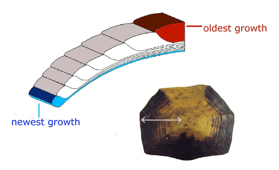
diagram of the supposed growth of laminae of the scute.
I have now photographed, measured and weighed the dead imported tortoises from Heathrow and have taken samples from the top (carapace) and underside (plastron) of the shell. It seems that the best tool to use is a dental scaler that has been bluntened slightly This gives more control over how much material is taken, which is important for two reasons: 1) If the technique is to be used on live tortoises it must be possible to do without fear of getting too near the nerve layer under the keratin, and 2) because I want to be able to get relatively good temporal resolution (see image).

areas sampled on plastron and carapace
As these are the first samples I’ve taken, I’ll be using them to check whether isotopic values are consistent within individuals. I therefore took samples from 4 different scutes on each tortoise (see image). I chose these scutes as I assumed they would be least susceptible to wear, though I’m not entirely sure I’ve got this right…
I also want to see if I can consistently sample a particular period of growth and have therefore sampled three areas of growth on each scute (following the growth rings). I am crossing my fingers on this one as it would be incredibly useful if I could sample multiple areas of growth to track changes in diet or location.
Next up: dissections!
(Image of scute cross section adapted from Seltzer and Berry (2005))
 RSS Feed

